Detector R&D and Applications in Biomedical Imaging
The group performs detector research and development (R&D), with possible spin-offs. The main activities concentrate around the upgrades of the CMS crystal calorimeter, together with R&D on radiation-hard scintillators. Most importantly, during the last decade the know-how on scintillating crystals and solid-state photo-detectors has led to the development of novel Positron Emission Tomography (PET) scanners for (bio-)medical imaging applications.
In the past the CMS ETH group had carried out studies on Lead Tungstate (PbWO4), the scintillator used for the CMS ECAL. These had shown that hadronic showers from energetic protons and pions cause a cumulative loss of light transmission in PbWO4. Following up on this, the group has performed extrapolations for the expected loss of light output in the CMS ECAL as a function of the integrated LHC luminosity. A further contribution by the group has been the identification and direct visualization of local centers of damage that might be due to fragments of the heavy elements, Pb and W, as the dominant cause of transmission losses. This qualitative understanding gave important guidance to further ongoing R&D studies of alternative radiation-hard scintillating materials (such as Cerium-Floride) for future calorimeters. Currently, in the context of the Phase-2 upgrades of the CMS detector for High-Luminosity LHC running, the group carries a leading role in the upgrade of the readout electronics of the CMS ECAL barrel part, including the detector control system, and contributes to the development and construction of the new precision timing layer.
The expertise on scintillating materials and solid-state photo detectors in the groups of Prof. Dissertori and Prof. Pauss was first used in a spin-off project (AX-PET), which proposed a demonstrator for a novel geometrical approach for high resolution and high sensitivity Positron Emission Tomography (PET) imaging, where a compact axial setup allows for a precise 3D localization of the photon interaction point in the detector, with no parallax error. Two AX-PET modules have been designed, built, commissioned and operated in coincidence, demonstrating competitive performance in terms of spatial and energy resolutions and delivering the prove of concept for this demonstrator, by the reconstruction of images of several phantoms.

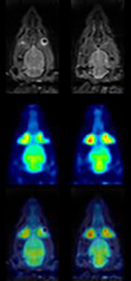
Then, a close collaboration with Prof. B. Weber (Univ. of Zurich) has been established and the SAFIR project was launched. SAFIR (Small Animal Fast Insert for mRi) is an innovative, high rate PET detector insert for MRI to be used for quantitative dynamic small animal imaging inside the bore of a commercial 7T MRI preclinical scanner operated by scientists from ETH and from the University Zurich, Institute of Pharmacology and Toxicology. The successful construction of a first two-ring prototype has shown that unprecedented temporal resolution (of a few seconds) and truly simultaneous PET/MR acquisition can be achieved. More details can be found on the SAFIR pages.
The expertise acquired thanks to the SAFIR project, together with the close collaboration with colleagues from the University and the University Hospital Zurich, was crucial for the recent design and development of a very cost-effective clinical brain PET scanner, for large-scale population scanning towards early diagnosis of neurodegenerative diseases. This has led to the founding of an ETH spin-off company (external page Positrigo AG), and the launch of a new SNSF-funded project, PETITION, with the aim to study further applications of this versatile and mobile brain PET scanner, in particular in the Intensive Care Unit (collaboration with the University Hospital Lausanne) and in proton-beam therapy (in collaboration with PSI).